Epiretinal Membrane
An epiretinal membrane is the growth of tissue over the center part of the retina, the macula. Although the exact cause is unknown, it is thought to be caused by normal cells that settle on the retina and grow in a thin sheet. This sheet may contract, resulting in wrinkling and swelling of the retina (macula edema).
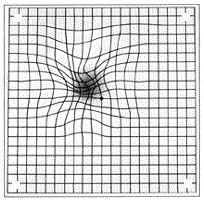
Most epiretinal membranes cause little or no symptoms and can be safely watched with serial exams. If an epiretinal membrane progresses this may result in blurring and/or distortion of central vision. These symptoms may be monitored at home with the use of an Amsler grid.
If symptoms progress to the point that patients are not able to perform their normal daily functions (reading, driving, etc) then surgery may be considered. Surgery consists of removing the gel from the back part of the eye (vitrectomy) with peeling of the membrane off the retinal surface.
The results of surgery are variable with most patients showing significant improvement in both blurred vision and distortion. The improvement may be seen after a few weeks and may continue to improve for up to a year or more.
Demographics
Epiretinal membranes typically occurs in those over age 50, and there is a slightly higher incidence in females. In most cases, the cause is unknown but predisposing conditions include retinal vascular disease (e.g. diabetic retinopathy), retinal tear, prior retinal detachment, and uveitis (ocular inflammation). The condition is mild in most cases and affects both eyes in approximately 25% of cases.
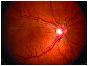
Appearance & Symptoms
Most epiretinal membranes are clinically insignificant and produce no symptoms or very mild visual disturbances. These membranes are often discovered on routine eye examination and present with a glistening or subtle sheen on the retinal surface. No treatment other than periodic monitoring is required, as approximately 70% of these will remain stable without intervention.
When an epiretinal membrane begins to cause symptoms, the membrane is more clearly visualized on the surface of the retina, and is sometimes called “macular pucker”. Definite wrinkling or striae of the retina are observed; the retinal blood vessels appear tortuous where the membrane is contracting and straightened or stretched in the surrounding areas. This produces symptoms of varying degrees of distortion depending upon the degree and location of traction induced by the epiretinal membrane. Careful examination will often reveal retinal swelling, or macular edema, which is responsible for blurred vision. Patients often describe their condition as ‘looking through a fish tank.’
Diagnosis
When an epiretinal membrane is discovered, certain diagnostic tests may be requested by your retinal specialist. The purpose of these tests is to evaluate a possible underlying cause of the epiretinal membrane, determine the degree of traction or macular edema caused by the membrane, and plan possible treatment.
The simplest technique is photographic documentation of the epiretinal membrane. Color and red-free photographs provide a valuable baseline for following the progress of membrane formation through time or evaluating a response to treatment.
Optical coherence tomography (OCT) is commonly used to obtain a high-resolution image of the epiretinal membrane and its effect on the retina. This technology uses laser light to scan the retina and construct an accurate cross-sectional image, non-invasively. The epiretinal membrane can be visualized along with its attachments to the retina, traction and distortion of the retinal architecture, and degree of macular edema. This is very helpful in guiding decisions for treatment.
For more information about this condition:
Epiretinal Membrane information from the ASRS Retinal Health series